CASE REPORT
Neonatal Cellulitis
R. El Qadiry1, 2, *, F. Bennaoui1, 2, N. El Idrissi Slitine1, 2, Fmr. Maoulainine1, 2
Article Information

Identifiers and Pagination:
Year: 2018Volume: 10
First Page: 142
Last Page: 146
Publisher ID: TOIDJ-10-142
DOI: 10.2174/1874279301810010142
Article History:
Received Date: 27/6/2018Revision Received Date: 17/9/2018
Acceptance Date: 24/9/2018
Electronic publication date: 17/10/2018
Collection year: 2018

open-access license: This is an open access article distributed under the terms of the Creative Commons Attribution 4.0 International Public License (CC-BY 4.0), a copy of which is available at: https://creativecommons.org/licenses/by/4.0/legalcode. This license permits unrestricted use, distribution, and reproduction in any medium, provided the original author and source are credited.
Abstract
Introduction:
Newborn cellulitis is rare and often atypical disease. It is a diagnostic and therapeutic emergency and its progress leads to instant death. We report three cases of neonatal cellulitis from the Neonatal ICU Department of Mohammed VI university hospital in Marrakesh, highlighting this rare, serious and unknown disease.
Case 1:
17-day-old female newborn admitted for sepsis and breast refusal since 5 days. Clinical examination revealed a hypotonic newborn, hypothermic at 34.2 °C with sclerema neonatorum. Skin examination found Erythema and edema on the left hemi-face with necrosis of the ipsilateral nostril. The blood culture had isolated coagulase-negative staphylococci. The newborn was put under triple antimicrobial therapy of 3rd Generation Cephalosporin (3GCs), gentamicin and vancomycin with good progress.
Case 2:
20-day-old male newborn, was circumcised ten days before admission, had sepsis with a fever at 40 °C evolving since 24h. The clinical examination found hard, hot and very painful inflammatory lesion starting at the scrotum and extending to suprapubic region and the start of the lower limbs. Blood tests revealed strongly positive infectious status. The progress was positive when put under triple antimicrobial therapy.
Case 3:
25-day-old female newborn, visited a traditional healer who gave her a mixture of unknown nature to apply on her face, hospitalized in our department for cellulitis of the right hemi-face extending to the periorbital and cervical region evolving since two days together with a fever. Infectious blood assessment was positive. The progress was favorable when put under triple antimicrobial therapy.
Conclusion:
Cellulitis is a rare disease in newborn and it must be diagnosed early because it can be complicated into sepsis. The search for other localizations, mainly meningeal, is essential.
1. INTRODUCTION
Cellulitis is an inflammation of the subcutaneous cellular tissue (the dermis) and it may extend to the surface or to the depth causing dermo-hypodermatitis. Newborn cellulite is rare and often atypical disease. Streptococci B group is the most convicted germ. It is a diagnostic and therapeutic emergency which leads to instant death [1-3].
The authors present three clinical cases of neonatal cellulitis and review its epidemiological, clinical and therapeutic characteristics, as well as its prognosis.
2. CASE 1
17-day-old female newborn was born prematurely at 32-week-pregnancy from a first-degree relative marriage, admitted for sepsis and breast refusal since 5 days.
Clinical examination revealed hypotonia, hypothermia at 34.2 °C with sclerema neonatorum. Skin examination found erythema and edema on the left hemi-face with necrosis of the ipsilateral nostril.
CBC showed: Leukocytosisat 12630 / mm3 predominantly neutrophilic 8530 / mm3 with lymphopenia at 1840 / mm3 and severe thrombocytopenia at 22 000 g / dl verified by peripheral blood smear. Plasma C-Reactive Protein (CRP) was 10.4 mg / L. The lumbar puncture found clear liquid with sterile culture. Two blood cultures were performed, one of which was done on the second day of antimicrobial therapy (3rd generation cephalosporin (3GCs) and gentamicin) because of the clinical worsening of the patient, a coagulase-negative staphylococci was isolated.
The newborn was then put under Vancomycin in addition to 3rd GCs and gentamicin for a period of 15 days, we noted a disappearing of the inflammatory lesion although the left nostril was amputated. Biological improvement was noted (leucocytosis at 11990 / mm3 with neutrophils at 3540 / mm3 and lymphocytes at 5070 / mm3 and a platelet level normalization at 140,000 / mm3).
3. CASE 2
20-day-old male newborn, was circumcised ten days before admission, had sepsis with a fever of 40 °C evolving since 24h.
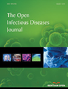
Clinical examination found a marbled newborn with cold extremities, tachycardia at 200 beats / minute, little reactivity, few gesticulations with plangent cries. Skin examination reveals hard, hot and very painful inflammatory lesion starting at the scrotum and extending to suprapubic region and the start of the lower limbs (Fig. 1).
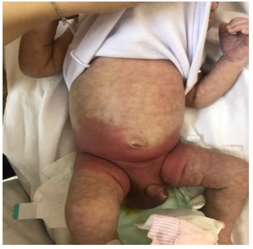 |
Fig. (1). Scrotal cellulitis with sepsis. |
Blood tests showed leukocytosis at 28,500 / ml predominantly neutrophilic at 15 530 / ml, lymphocytes at 8260 / mm3 and platelets at 408,000 / mm3, CRP at 204 mg / l. In addition to that, the two blood cultures made during febrile peaks, cerebrospinal fluid and CBEU were sterile. Scrotal ultrasonography, without skin biopsy, had noted a thickening with infiltration of the scrotal walls extended to the perineal region and the abdominal wall without signs of dermo-hypodermitis such as abscess collections.
The progress was positive when put during 15 days under triple antimicrobial therapy (amoxicillin, 3rd generation cephalosporin and aminoglycoside) by achieving apyrexia after 48 hours of treatment and disappearance of local signs without any scars at the end of treatment (Fig. 2).
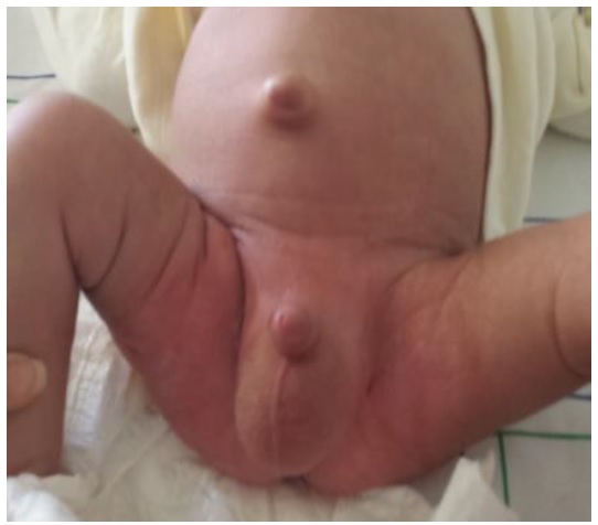 |
Fig. (2). Scrotal cellulitis after the treatment. |
4. CASE 3
25-day-old female newborn, had visited a traditional healer who gave her mixture of unknown components to be applied on her face for cosmetic reasons. She was admitted two days later for an inflammatory lesion of the right hemi-face extending to the periorbital and cervical region (Fig. 3) with a fever. The clinical examination revealed an irritable newborn, tachycardic at 180 beats / minute with signs of sepsis and erythematous, hot and very painful inflammatory lesion, extended from the right hemi-face, to the neck and to the upper part of the thorax. There was also edema of the right eyelid, making it difficult to examine the eyeball.
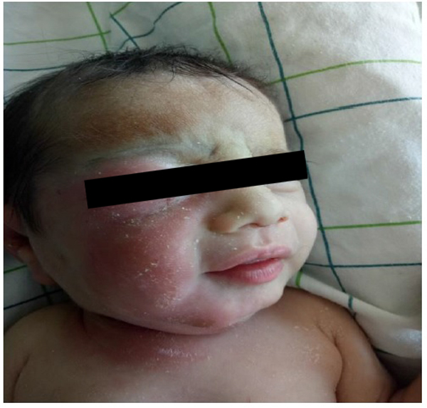 |
Fig. (3). Orbito-facial cellulitis before treatment. |
The orbito-facial CT-scan showed subcutaneous fat densification of the right jaw region and presteptal fat of the right orbit without detectable collection.
The biological assessment had shown a very high CRP at 290 mg/l and leucocytosis at 17620 / mm3 predominantly neutrophilic at 12281 / mm3 with lymphocyte count at 4581 / mm3 and platelets at 221000 / mm3. The lumbar puncture and the only blood culture performed returned sterile.
The progress of the disease was positive under triple antimicrobial therapy based on amoxicillin, 3GCs and gentamicin, with decreasing of the inflammatory lesion and normalization of the biological tests (the CRP at 1.12 mg / l, the leucocytosis at 13810 / mm with neutrophil at 5930 / mm3 and lymphocytes at 6030 / mm3) after 15 days of treatment (Fig. 4).
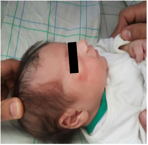 |
Fig. (4). Orbito-facial cellulitis after the treatment. |
5. DISCUSSION
Cellulitis in newborns was described for the first time in 1980 under the term “cellulitis-adenitis syndrome”. It is extremely rare and the data on this atypical manifestation are, however, very limited and are mainly based on small series or isolated observations [1].
Group B Streptococci (GBS) cellulitis is the most commonly described. They seem to be increasing and it is recommended in front cellulitis of newborn and infant less than 3 months to introduce in first line of treatment an antimicrobial active on the GBS, while respecting the recommendations of the materno-fetal infections.
There are also cases where authors described a Methicillin-resistant Staphylococcus aureus, a group A streptococcus or an E. coli, hence the need for empiric antimicrobial therapy, to be adapted later according to biological findings [3, 4]. In our patients, we did not isolate any germ; this could be explained by the limited number of blood cultures carried out.
The infectious invasion is often hematogenic during a bacteremia, but sometimes if it has cutaneous entryas the case of our patient or by contiguity during a ethmoiditis or a dacryocystite.
The initial symptoms of cellulitis are mostly non specific signs such as fever, irritability and digestive disorders. Some studies have reported cases of infants initially having isolated swelling without systemic signs [3]. The diagnosis must be made early because it is often combined with bacteremia and meningitis and the search for meningeal location is generally recommended even in asymptomatic newborns.
The most frequently described locations are sub-mandibular [5], or more rarely retroauricular, inguinal or scrotal [1].
The treatment is based on empiric antimicrobial therapy combining a 3rd generation cephalosporin (cefotaxime) and an aminoglycoside (gentamycin). In case of associated hemodynamic disorders suggesting invasive infection with Staphylococcus aureus or Group A streptococci, an antimicrobial with anti-toxin activity [1] is added. The period of treatment is different from one study to another. It should be guided by the clinical response and the progress of biological inflammatory assessment especially C-reactive protein but given the risk of relapse, intravenous treatment should be extended at least 10 to 14 days [3].
CONCLUSION
Cellulitis is rare in newborns but its progress is quick and lethal. In order not to misunderstand severe sepsis and to optimize management, early diagnosis is important. The search for other localizations, particularly meningeal, must be systematic.
ETHICS APPROVAL AND CONSENT TO PARTICIPATE
Not applicable.
HUMAN AND ANIMAL RIGHTS
No animals/humans were used for studies that are the basis of this report.
CONSENT FOR PUBLICATION
A written informed consent was obtained from the parents.
CONFLICT OF INTEREST
The authors declare no conflict of interest, financial or otherwise.
ACKNOWLEDGEMENTS
Declared none.